시반이 이동되지 않는 이유 (시반의 고정, 양측성 시반, 고정 시반)
시반의 변위가 일어나지 않거나 시반의 이동이 없는 것은 이제까지는 자가용혈로 인한 헤모글로빈의 조기 확산 때문이라고 알려져 왔으나
사실은 힐거만의 이론대로 유체정역학(流體靜力學) 압력과 관련된 것으로 혈관 벽을 통과하는 혈액내의 액체 손실로 인한 농축(hemoconcentration) 때문이다.
The reason for non-displacement and non-shifting of lividity is not the early diffusion of haemoglobin, as supposed formerly, but the haemoconcentration by loss of fluid which penetrates the wall of those vessels related to the hydrostatic pressure, as shown by Hilgermann.
The Estimation of the Time Since Death in the Early Postmortem Period, 1995, Edward Arnold; C Henssge, B Knight, T Krompecher; pp241
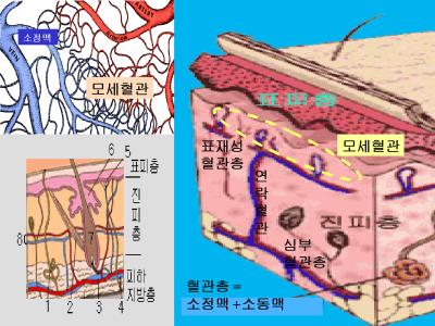
일본의 Noriko 교수는 시반의 고정 매커니즘을 알기 위해 면역조직학적 연구를 했다. 사망 후 7.5 시간부터 2주까지의 68구 사체에서 피부 조직을 검사하였다. 혈색소가 침투된 피부 조직의 여러 부위별 (예를 들면 혈관내 적혈구, 혈관벽, 혈관주위조직, 진피결합조직) 검사를 시행했다. 지압흔이 관찰되는 곳에서는 49%가 고정시반이 있는 곳에서 41%에서만 혈색소의 피부조직 확산이 관찰되었다. 이러한 소견은 시반의 고정이 혈색소의 확산 때문이 아니라 혈액의 농축 때문이라는 가설을 뒷받침해준다.
An immunohistochemical investigation of postmortem lividity was performed to illuminate localization of hemoglobin (Hb) and the mechanism of fixed lividity. The fixed lividity was defined as an unfading phenomenon by thumb finger pressure. Skin specimens were taken from 68 autopsy cases 7.5-336 h (2 weeks) postmortem. Localization of Hb of the specimens was examined by a labeled streptabidin biotin (LSAB) method using polyclonal (rabbit antihuman hemoglobin antibody) and monoclonal (mouse anti-human hemoglobin monoclonal antibody) antibodies. Positive staining for Hb was observed in various sites of the skin, i.e. in only intravascular erythrocytes, in vascular walls and perivascular tissue including sweat glands and sebaceous glands, in the dermal connective tissue, and in almost all of skin tissue except the horny layer. The diffusion of Hb into skin tissue was observed in 20 of 41 displaceable lividity cases (49%) and 11 of 27 fixed lividity cases (41%). Compared to displaceable lividity, superficial plexi in fixed lividity were filled with erythrocytes, which were markedly immunodetected. These findings support the hypothesis that the fixation of lividity is not due to diffusion of Hb into skin tissue but hemoconcentration in blood vessels. Forensic Sci Int 1995 Apr 27;72(3):179-89
Penttila와 Laiho는 사체의 말초 혈액에서 다양한 형태의 세포 형태를 분석하였다. 그 결과는 다양한 혈액 세포의 구조물이 자가용혈에 상당한 저항성이 있는 것으로 나타났다. The Estimation of the Time Since Death in the Early Postmortem Period, 1995, Edward Arnold; C Henssge, B Knight, T Krompecher; pp224
(참조 1)
1. 부패되기 시작하면 적혈구가 깨지면서 나온 헤모글로빈이 혈관벽을 빠져 나와 조직에 침착됩니다. 과거에는 양측성 시반이나 고정 시반이 생기는 원리를 모두 이것으로 설명했었습니다.
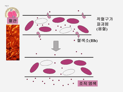
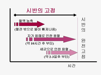
2. 혈액은 적혈구가 약 45%이며 일부 백혈구와 기타 고형성분을 빼도 약 50%가 물입니다. 사망후 이 물이 먼저 혈관벽을 빠져나가면서 농축이 된다는 것입니다. 농축이 되면 될수록 혈액의 이동은 방해받게 됩니다.

(참조 2)
Older theories about 'fixation' of the staining after a certain time are not tenable, as there is no constant interval when this occurs. Formerly, it was held that if a body had remained in its original posture for a certain minimum length of time the blood would coagulate in the hypostatic areas, so that secondary shifting could not then occur if the body was moved. This is not true in the majority of instances, as there may be partial or complete secondary gravitation at any time- at least, until true staining of the tissue due to haemolysis begins as part of early decomposition, which is not until the second or third day in temperate conditions. Bernard Knight, Forensic Pathology, pp. 53
It may offer some indication as to whether the position of a body has been altered after it has become fixed(about 4 to 8 hours). Fracis E. Camps, Practical forensic medicine, pp.104
After 8 to 12 hours lividity becomes "fixed" and will remain where it originally formed. Simpson and Knight, Forensic Medicine pp. 9
After about 10-12 hours the lividity becomes "fixed" and repositioning the body, e.g. from the prone to the supine position, will result in a dual pattern of lividity since the primary distribution will not fade completely. Fixation of lividity is a relative, rather than an absolute, phenomenon, but nevertheless, well developed lividity fades very slowly and only incompletely. Duality of the distibution of lividity is important because it shows that the body had been moved after death. However, the timing of this movement of the body is inexact. Prof. Derrick J. Pounder.
After 4-5 days the ever-increasing haemolysis in the vessels produces sufficent pigment to stain the hypostatic areas, and this is permanent. Gradwohl's legal medicine pp.81
(참조 3)
Autolytic changes in blood cells of human cadavers. II. Morphological studies. The morphology of various types of cells in the peripheral blood of human cadavers was investigated. The material comprised 123 medicolegal autopsy cases with post-mortem (p.m.) times ranging fro 1.7 to 270.4 hours. The corpses were kept at +4 degrees C. The haematocrit values of the blood increased rapidly after death. The haematocrit-corrected red cell count, and the total white cell and platelet counts remained quite stable during the whole p.m. time range. Red cells were quite rapidly transformed from a discoid configuration to crumbled discs, echinocytes and spherocytes, but no debris or burst cell configurations were seen. Rapid deterioration of the staining properties and marked morphological changes in many leucocytes occurred quite rapidly after death. Lymphocytes seemed to be the most resistant and basophils the least resistant to the effects of autolysis. Morphologically altered platelets and aggregates of them were seen in each cadaver. The present morphological observations and the quantitative results suggest that various cellular elements of the blood seem to be quite resistant to autolytic effects, and many cells apparently retain their viability for longer periods of time in the blood of cadavers kept at reduced temperature. Penttila A, Laiho K.; Forensic Sci Int 1981 Mar-Apr;17(2):121-32

